Latest Bone & Joint Articles | Wockhardt Hospitals

ARTICLES ON Bone & Joint

Latest Articles on Bone & Joint

Makoplasty - Robotic Knee Resurfing

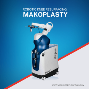
Makoplasty : Robotic Knee Resurfacing

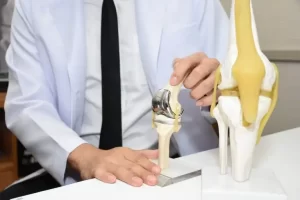
Dr. Mudit Khanna at Wockhardt Hospitals, Mumbai Central has pioneered the use of Mako SmartRobotics in South Mumbai to treat arthritis of the Knee. He is the first surgeon in the region to perform Makoplasty Robotic Knee Resurfacing and has now gained significant experience with this technique. Makoplasty: An Alternative to Total Knee Replacement Makoplasty Robotic knee resurfacing can be an attractive alternative to total knee replacement in the appropriate patient. This technique, which involves robotic bone contouring of arthritic surfaces and customized resurfacing of the damaged area, represents an improvement over traditional or conventional partial (or unicondylar) knee replacement. Without robotic assistance, partial knee replacement can provide excellent and durable pain relief but is technically challenging, and carries a higher potential for surgical inaccuracy. Dr Mudit Khanna’s experience, combined with Mako SmartRobotic safeguards, provide an unparalleled level of accuracy, precision and personalization to optimize patient outcomes. Advantages of Robotic Assistance in Knee Resurfacing After robotic knee resurfacing, Dr. Khanna expects patients to return to work much faster. Compared to total knee replacement, robotic knee resurfacing provides more reliable return to select sports, a quicker and less painful recovery, a smaller incision, a more natural feeling knee, and, importantly, a lower risk of complications. Dr. Khanna performs all his Makoplasty knee resurfacing procedures at the SOBO Unit of Wockhardt Hospitals which carries one of the lowest infection rates in the region. Comprehensive Pain Management and Rehabilitation Realizing the significance of Early rehabilitation and OLXGG of Post-operative Pain management, Dr. Mudit Khanna with the help of the department of Anesthesia has established an excellent Enhanced Recovery After Surgery (ERAS) Program at Wockhardt Hospitals, which helps to achieve Rapid recovery after Joint Replacement Surgery by improving the quality of perioperative care with special focus on achieving excellent pain management through a multimodal pain management protocol. In addition to Dr. Mudit Khanna’s expertise, the world-class environment at Wockhardt enhances the patient experience and outcome. Schedule your consultation with Dr. Mudit Khanna today and take the first step towards a pain-free, active life with Makoplasty Robotic Knee Resurfacing!

Read More
avascular necrosis of femoral head

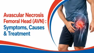
Avascular Necrosis Femoral Head (AVN) : Symptoms, Causes & Treatment

It’s easy to shrug off a dull, deep ache in your groin as just a pulled muscle, especially if you only notice it while climbing stairs. But for many, that quiet discomfort is actually the first warning sign of a serious bone issue that only gets tougher to handle the longer you wait. Avascular necrosis of the femoral head, also called “osteonecrosis”, happens when the top of the thigh bone loses its blood supply and slowly begins to die. It continuously strikes the hip joint. If it is caught early, the damage can be slowed down, but if it is ignored, the bone can dig in and wear out the whole joint. This blog explains the warning signs of AVN, what sets the condition off, how doctors find it, and the treatment choices available. What Is Avascular Necrosis of the Femoral Head? Bone is a living tissue, and it needs a steady flow of blood to stay healthy. Avascular necrosis (AVN) is what happens when that flow is cut off, and part of the bone starts to die. The name is easier than it looks. The “A” stands for without, while “vascular” means blood vessels, and “necrosis” means the death of the tissue. Doctors also use the word osteonecrosis, which just means bone death. Without the supply of fresh blood, tiny cracks can start to form inside the bones. Over months, the weakened area can begin to collapse.  Most cases appear between 30 and 50 years old people. In India alone, doctors diagnose an estimated 80,000 to 90,000 new cases every year. Anyone can get affected by it, which is why the early signs are worth knowing. Symptoms of Avascular Necrosis of the Femoral Head Early avascular necrosis symptoms can be silent. Plenty of people feel nothing while the damage is small. As the bone weakens over time, many people start feeling the pain. At first it can show up when the joint carries weight, and later it can start nagging even during rest. Pain from a bad hip is often first felt in the groin area. Some people notice it in the thigh or the buttock instead. The most common signs are: Mild, nagging pain that can get worse with time. Joint getting stiff, making it harder to move around like you usually do. Pain that spreads from one spot to other areas. Struggle standing up or walking for long. Muscles around the joint that grow weak and shrink from being used less. In severe cases, the bone can collapse and bring sharp pain. Pain that lingers for more than a few weeks, or that wakes a person at night, is a solid reason to have the hip checked. Causes of Avascular Necrosis of the Femoral Head Blood reaches the femoral head through a small set of vessels. If there is anything that blocks or damages these vessels, it can starve the bone. Several injuries and health problems can cause avascular necrosis of the femoral head. This can include: Injury or Trauma: A hip dislocation or a broken bone can tear the vessels that supply blood to the femoral head. Steroid Medicine: Long-term or high-dose steroids, often given for asthma, arthritis, or immune conditions, can raise the risk by changing how blood vessels work. Heavy Alcohol Use: Drinking a lot over many years can clog vessels with fat and weaken bone, especially around the hip. Certain Medical Treatments: Radiations given for cancer can also harm bones. Many organ transplants, like a kidney transplant, are linked to causing AVN. Bisphosphonates: This is a bone-strengthening drug, and in rare cases can cause bone death in the jaw. The risk is higher in people treated for multiple myeloma or breast cancer. AVN that is not caused by an injury is tied to several other conditions. Among them are:  Diabetes HIV Sickle cell disease Pancreatitis (swelling of the pancreas) Gaucher disease (a build-up of fat inside the organs) Decompression sickness (gas bubbles in the blood from fast pressure changes) In more than half of people, both hips are affected, so a problem on one side is a signal to keep an eye on the other. How Is Avascular Necrosis of the Femoral Head Diagnosed? Getting AVN diagnosed early can give a real chance to save the joint. A doctor usually starts with questions and a hands-on check, then confirms the diagnosis with imaging. Medical history and physical exams usually come first. Your doctor will then ask about the pain, any past injuries you had, or medications you have been taking.  To see how much damage there is, doctors rely on imaging tests: An X-ray is often the first step, as it can clearly show if the bone’s shape has changed or if the joint is starting to collapse. Magnetic resonance imaging (MRI) can give detailed pictures and can catch AVN in its very first stage.  A computed tomography (CT) scan can offer cross-section views which can help map out the exact bone damage done. Bone scans act as a small, safe tracer that highlights areas where blood flow might be restricted. Biopsy is usually the last option, where a small piece of bone is checked under a microscope. Understanding the Stages of AVN of the Femoral Head AVN gets worse in stages. Spotting it in an early stage can make the treatment far more successful.  Early Stage: At this stage, the blood flow to the femoral head drops and bone cells begin to suffer. The bone can still hold its shape, and there may be no pain yet. An MRI can pick up these changes easily. Bone Death Stage: This is the second stage, where the blood flow is still cut off, and now the bone tissue starts dying. Pain often starts at this stage, mostly when the weight falls on that weakened hip. Here, rest and medicine are advised by doctors. Crescent Stage: In this stage, dead bone weakens, and a small crescent-shaped crack starts to form under the surface. The

Read More
electric buzzing feeling in body

Electric Buzzing Feeling in Body: Feet, Toes, Hands, Fingers, Arms, and Legs: Anxiety

Have you ever lain in bed at night and felt a weird, faint vibration under your skin? You look, but there’s nothing you can see, no twitching muscles or shaking hands. It’s just this strange sensation, like your body has a motor running that won’t power down. It feels almost exactly like having a phone vibrating under a cushion, except the vibration is coming from inside you. Doctors commonly call this feeling an internal tremor, or an internal vibration. A person can feel it in the feet, toes, hands, fingers, arms, legs, or the whole body at once. Sometimes it can come and go, while other times it can stay there for weeks. It can sometimes sit at one spot, and sometimes it can travel all across the body. Most of the time it happens on its own, without any warning. The feeling is real, and it is quite common. What makes it grow is worrying about it too much. A quiet buzz can turn into a loud one once a person starts to fear what it actually means. This blog goes through the common causes of this electric buzzing feeling in the body, how anxiety can feed it, the warning signs to check for, and simple steps that help calm it down. What Does an Electric Buzzing Feeling in the Body Feel Like? People use many words for the same thing. Some describe it as fizzing, some say humming, some call it a tremble deep under the skin. A few say it feels like sitting on something that keeps buzzing. Your hand might look completely still on the outside, even though you can feel it buzzing deep down. During the day, work and noise can pull attention away from the body, but at night the buzzing can usually get louder because the body is at rest. If you are tired, the nerves can get even more touchy. A long, stressful day can give strong vibrations. Many people report that the feeling is worse just as they try to fall asleep. The location can move too. One night you can feel it in the legs, another day it can hum in the chest or belly. Knowing that the sensation can shift and still be harmless can take some of the fear out of it. What Causes the Buzzing Feeling in Feet, Hands, Toes, and Fingers? An electric buzzing feeling can come from many places, from harmless and short-lived to signs of something that needs care. A tired, stressed, or slightly out-of-balance body sends odd signals along its nerves, and buzzing is one of them. Here are the usual reasons. Nerve irritation: A squeezed, swollen, or irritated nerve is one of the main reasons behind the buzz. The pressure or inflammation felt on the nerve can send strange signals that can almost feel like electric vibrations. Neurological conditions: If there is damage to the nerves, this can cause buzzing in the body. Different conditions like multiple sclerosis (MS), diabetic neuropathy, and peripheral neuropathy can also cause internal tremors.  Anxiety or stress: If a body is under too much stress, it can also cause the same buzzing feeling. High anxiety can switch the nervous system into a fast, alert state, and that extra activity can show up as numbness, tingling, or a buzzing feeling in the body. Pinched nerves: Pressure on a nerve can cause a shock-like feeling. Bad posture or a spine slightly out of line can all pinch a nerve. Poor blood flow: Nerves need a steady supply of blood. Changes in circulation, sometimes linked to conditions like peripheral artery disease, can starve a nerve and set off an electric buzzing feeling in the body. Medication side effects: Medicines like antidepressants and anxiety medicines can also cause a buzz in the hands or feet. Too little sleep: Not having enough rest can make the body more sensitive to the buzzing. Poor sleep raises cortisol, a stress hormone that keeps the nervous system switched on and vibrating. Hormone shifts: Muscles and nerves work according to the hormones following in our body. An overactive thyroid, the swings of perimenopause, or the days before a period can each cause trembling vibration in the body. A dip in blood sugar can also leave a person buzzing.  Too much caffeine: Drinking too much coffee, tea, energy drinks, or even taking certain medications can kick your nervous system into overdrive and might start that jittery, humming sensation. Low vitamins or minerals: If the body has a shortage of vitamin B12, vitamin D, or magnesium, this can impact nerves and cause vibrations. Even very high or very low levels of body salts can cause vibrations. Understanding the reason behind this electric buzzing in the body is important because the fix depends on it. Can Anxiety Cause the Internal Vibrations? Stress and the cause of vibrations are closely tied together. When the brain senses danger, even if it’s not real, it flips into a fight-or-flight mode. The heart rate starts climbing, breathing speeds up, and muscles start to tense and load with energy, making the body ready to just run from the situation. This primed state can feel like a hum running through the body. Experiencing stress for a long period of time keeps the body stuck in flight-or-fight mode. When your nervous system is constantly on high alert for weeks, it can start sending out vibrations even when your body feels calm. Researchers are still studying exactly how this buzzing in the body works, and most of them point to how hormones react to long-term stress. It’s tempting to search for your symptoms online, but this can often lead you to health sites discussing rare conditions like MS and amyotrophic lateral sclerosis (ALS), which can in turn increase the fear that follows and tighten the body even more. More fear can create more buzzing, which can lead to more searching. Breaking that loop is often the first real step to get some relief. Is The Internal

Read More

Bone health tips: Expert on ways to get enough vitamin D for healthy bones

A large number of people are known to be deficient in vitamin D and face a plethora of health issues. Vitamin D deficiency is commonly associated with bone and muscle pain. Here’s how to get the right amount of it to maintain a good bone mineral density in people of all age groups. Vitamin D, also known as the sunshine vitamin is necessary for one’s overall well-being but did you know vitamin D helps to regulate calcium and phosphate in the body and improve your overall well-being? Moreover, enough vitamin D is also necessary for healthy bones and since it supports your bone health, try to include enough amount of this sunshine vitamin in your diet to stay fit and fine throughout life. Currently, a large number of people are known to be deficient in vitamin D and face a plethora of health issues since there is not much awareness regarding this sunshine vitamin and people fail to understand what exactly its role is. In an interview with HT Lifestyle, Dr. Pankaj Gunjal, Consultant Orthopedic Surgeon at Wockhardt Hospitals in Nashik, shared, “A good amount of vitamin D is needed in order to prevent metabolic syndromes, diabetes, autoimmune diseases, cardiovascular diseases, respiratory infections, food allergies, and asthma.” He added, “Vitamin D deficiency is commonly associated with bone and muscle pain. You will be shocked to know that being deficient in vitamin D can lead to skeletal diseases. The right amount of it can help to maintain a good bone mineral density in people of all age groups.” Decoding the connection between vitamin D and bone health, he said, “Vitamin D aids in strong bones and muscles. Without the recommended amount of Vitamin D, one will not be able to absorb the calcium that is required for good bone health.” He cautioned, “Those children who are deficient in this vitamin can suffer from a condition called rickets leading to bone weakness, bowed legs, and other skeletal deformities like stooped posture. One can also have problems such as osteomalacia which is softening of bones, fractures, aches, and pains. Vitamin D decreases bone loss and reduces the risk of fracture in older men and women. So, along with calcium, vitamin D helps to keep osteoporosis at bay. To absorb calcium in the correct way, an adequate amount of vitamin D must be present in the body.” Suggesting how to get enough amount of Vitamin D, he advised, “It will be imperative for you to get enough amount of sunlight on a daily basis. Yes, that’s right! Try to expose yourself to the sun for at least 15-20 minutes between 10:30 am to 01:30 pm. Remember, overexposing can damage the skin so be careful. Make sure to include vitamin D-rich foods in the diet. Eat mushrooms, egg yolks, salmon, mackerel, and tuna. Take vitamin D supplements only after the doctor’s advice. Take a vitamin D3 test from time to time to check it and ensure that you stay hale and hearty. Also, avoid going overboard on vitamin D as high levels of it lead to nausea, vomiting, and weakness.” Dr. Pankaj GunjalConsultant Orthopedic SurgeonWockhardt Hospitals, Nashik To book an appointment call: +918669998260 Source: https://www.hindustantimes.com/lifestyle/health/bone-health-tips-expert-on-ways-to-get-enough-vitamin-d-for-healthy-bones-101671524010364.html

Read More

Tips to enjoy a healthy pain-free ‘Shoulder’ Joint

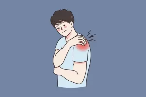
In everyday life, it’s your shoulder doing all the work. It has to move in everything that you do. You use it for lifting, reaching out to pick up something, reaching overhead to get that book off the shelf, or even pitching a ball and swinging your bat.  You can do all of these activities since you have a supple, flexible shoulder joint with pristine cartilage and all the surrounding muscles working in rhythm. But everything that moves around so much and is like a “golf ball on a tee” is also more prone to get injured. You will be surprised to know that the shoulder joint is the body’s most injured joint. It’s the repetitive motion of the shoulder joint (especially strenuous overhead motion) for someone involved in activities like painting, hanging curtains, working as a hairdresser, and lifting heavy weights in a gym that takes a toll on this otherwise forgiving joint.  Your shoulder can get hurt in other ways too: Prevention The great news is that most of these shoulder pathologies are easily treatable without surgery.  Of course, ‘prevention is better than cure. Some simple ways to avoid shoulder pain are- Dr. Mohit M KukrejaConsultant – Orthopedic SurgeonWockhardt Hospitals, Mumbai Central

Read More

Do You Feel Pain in Knees While Climbing a Flight of Stairs?

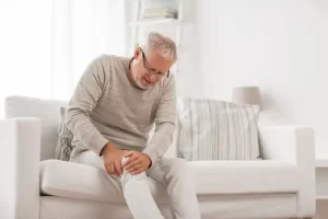
Have you experienced a time when every time you stood at the bottom of the steps and looked up thinking it was a physical endurance test to climb those stairs? Each step causes excruciating pain or extreme discomfort in the knees no matter how slowly or carefully you climb. The pain, which started in his right knee, now affected his left? Has knee pain ever caused you or a loved one to avoid activities? The reasons could be many. It may be due to damaged cartilage in your knee. The damage could be reversible (chondromalacia) or irreversible (arthritic) Why Does Your Knee Hurt When You Climb the Stairs? To answer that question, let’s review the mechanics of knee movement. Your knee joins three major bones: your thigh bone (femur), your shin bone (tibia), and your kneecap (patella). All three bones along with their associated ligaments and muscles must work in unison to support your weight and allow fluid leg movement. With each bend, the patella, a free-floating bone, slides over the femur in the trochlear groove. The articular cartilage keeps your kneecap in position as it cushions and lubricates the joint, so the bones glide against one another. Damaged cartilage may not cause pain when you walk, especially if it affects the knee cap cartilage in isolation. Running, deep knee bends, squats or climbing stairs subjects the knee to additional stress and the pain increases. These types of motions force the kneecap to slide up and down. Worn cartilage cannot keep the kneecap in the groove when the knee is under pressure. As the kneecap slips out of position, it causes pain. How is Knee Cartilage Damaged? Fractured bones or injured muscles can cause an imbalance of strength in the leg and pull the kneecap to one side of the groove or another. The added stress can cause misalignment and pain. Overuse injuries cause damage to the cartilage, especially in young athletes. Growing bones and excessive stress create a recipe for a chronic condition. Prolonged knee pain after activity indicates a more serious condition than normal muscle soreness from a strenuous workout. If you experience knee pain after working out, playing sports, or any strenuous activity that does not diminish in 72 hours, you may need medical attention. Genetic deformities and age are risk factors that cannot be modified but there are some things you can do to avoid the progression of your symptoms or delay damaging the cartilage. What is the Most Appropriate Treatment for a Runner’s Knee? The best way to avoid chondromalacia is to maintain the correct alignment of the kneecap and minimize wear and tear of the cartilage. Step 1: Develop strong and balanced leg muscles above and below the knee. These muscles support and stabilize the patella to keep it in the trochlear groove. Please remember that the muscles at the front of the leg act in opposition against the hamstrings to bend and straighten the knee. As one muscle contracts or gets shorter, the other muscle lengthens. An imbalance of strength can pull the kneecap out of position. Step 2: Assess your posture and the alignment of your feet and legs. Here are three tips to remember. Repeat the steps for each stair you climb. If your knee pain increases or lasts for more than two weeks, it’s time to contact your orthopedic specialist. Please note – Continuing to walk on a sore knee can cause significant internal damage to the tendons and ligaments inside your knee. Dr. Mohit M KukrejaConsultant – Orthopedic SurgeonWockhardt Hospitals, Mumbai Central

Read More

Joint Replacement Way to Mobile Life

Everyone will have arthritis in their old age and post-menopausal woman may have arthritis at an early age. For the last 12-13 years, I am doing knee replacements in which I found that out of my total number of cases more than 60-70 % are female patients and usually they get operated on at the age of 60-70 years and males usually get operated in their 70-80s. Knee Replacement is rewarding surgery for those who really get disabled and should be get done at proper time not too early neither too late to keep you active in old age as it improves your activity and overall health. It gives you painless, mobile, and stable joints. A surgeon’s proper training and experience are a very important factor in deciding the best outcome of this surgery.Also truly speaking neither any advanced gadget like robot or navigation will give good result in inexperienced hands and it’s debatable nowadays all those fancy gadgets really required for experienced surgeons as long term outcome depends on a lot of other factors like age of the patient, associated medical conditions, obesity, cementing technique, bone quality, etc. Unless surgeon knows properly what is knee replacement he can’t use it and one who knows properly may doesn’t require it.Also, long-term results are yet to be proved. Usually, the patient is mobilized on the next day and within 3-4 days patient becomes comfortable walking with a walker and using the western toilet. Even stairs climbing can be started on the 5th day.By 2-3 weeks, the patient starts walking without support. This surgery gives dramatic results for those who are disabled due to knee pain.

Read More

Joint Replacements of the Knee, Hip, Shoulder and Elbow are Life-Changing, Long-Lasting Solutions

While past generations often put off seeing a doctor for joint pain, today’s active adults are finding relief with total joint replacement surgery. Wear and tear on joints are a natural part of aging, so too are the inevitable aches and pains that come with it. Osteoarthritis is the most common type of arthritis, affecting more than 56.6% elderly population in India. The bones that make up your joint surfaces are covered with a protective layer of cartilage that absorbs shock and vibration and allows the bones to glide smoothly during motion. Arthritis causes the cartilage to gradually wear away, resulting in a painful rubbing of bone-on-bone. All of your joints may be affected by osteoarthritis, although weight-bearing joints, such as the hip and knee, tend to be the most painful. Other joints such as the shoulder and elbow are also involved causing annoying pain and functional disability. People who cannot function day to day because of pain or having rest or night pains and have restricted mobility are good candidates for surgery. The main objective of joint replacement surgery is to replace the injured or wound joint surfaces with materials such as metal, plastic, polyethylene, ceramics, or a combination of materials. The material selected depends on the reason for the surgery, as well as which joint is being replaced. There are more varieties of artificial joints available now than ever before and orthopedic surgeons will help decide which option is the best for your individual needs. Results after joint replacement are more consistent now thanks to new techniques, technologies, and materials. Few knee replacements in selected patients can be done minimally invasive subvastus approach having faster recovery, short stay, and less pain. Also, there is no absolute age restriction for these surgeries with average ages of 50 to 80years. It is proved now that joint replacement tends to be easier when you prepare for it like weight reduction, preoperative exercise protocols, improve safety and comfort at your home for easier postoperative independent ambulation. Knee replacement is the most successful surgery in all of medicine and most people have a better quality of life. Hip replacement is also a very successful common surgery done by orthopedic surgeons mostly for avascular necrosis, hip fractures, and hip arthritis and making most patients ambulatory the second day after surgery. Patients with shoulder and elbow arthritis problems try to bear and tend to delay or avoid joint replacement for long. But in the last two decades with advancements in shoulder and elbow replacement, we are able to give predictable best functional outcomes. Patients should be encouraged for these surgeries to lead pain-free and functional daily activities of the upper limb. Joint replacement recovery can be long about 6 to 12 weeks and involves a lot of walking, daily exercises and rehabilitation program. Your new joint may feel or sound different, which is normal, but you will get tuned to it with time. Gradually you will have full recovery and can look forward to returning to active life and leaving pain behind. Joint replacement cost is often a concern for people considering surgery. Most health insurance plans cover this surgery and also many government schemes can be availed. Considering high burden of disease even package prices have come down considerably to make it more affordable. Joint replacement implants have mechanical components, which have wear and tear with them. However, more than 90%implants will last 15years at least. Some will last much longer. You can extend the life of your implant by maintaining a healthy weight, avoiding high-impact activities, and staying fit and active. It is essential to have regular physiotherapy and orthopedic surgeon follow up to monitor implants for longevity. Patient awareness, education, improved healthcare facilities, gadgets, technological advances, implant inventory, improved pain management, and anesthesia practices have certainly made joint replacement a big boon to humans suffering from arthritis.

Read More

Newest Advancement in Joint Surgery – ARTHROSCOPY

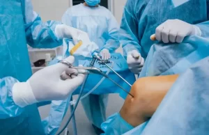
Arthroscopy is a surgical procedure performed on the joints by a special instrument called an Arthroscope and has developed into a new and reliable technique in the field of medicine. There is not much awareness in society about it. In the last few years, radical changes have taken place in the medical field. Earlier, major surgery was required for the disorders, it was a combination of a large incision, excessive bleeding, longer hospital stay, and longer recovery time after surgery. But today, it is not like that. Due to the progress in the medical field, it has become possible to perform surgery without any major incision, as a result, the patient can recover faster and start his daily routine early. Let us now understand what Arthroscopy is and how it is done using an Arthroscope. Arthroscopy is a surgery performed on Joints with help of a special device known as an Arthroscope, which is only 4 mm wide. This device allows the doctor to see the structure of the joints clearly on a monitor screen and make it possible to operate on the joint without making a large incision. Knee, shoulder, hip, elbow, wrist, and ankle joints can all be operated with arthroscopy. If the patient has any complaints like cartilage tear, ligament tear, injury to cartilage, knee arthritis, injury to the knee cap, frozen shoulder, rotator cuff tear, frequent shoulder dislocation, all ailments can be treated by arthroscopy. Treatment is definitely beneficial. There is no need to be afraid of surgery at all, be it is a sports injury, arthritis, or an accidental joint injury, due to technological improvement it is effortless to operate on joints without any incision through Arthroscopy. This results in less bleed during the surgery, less pain, and the patient gets discharged from the hospital early, gets recover faster, and can start his daily routine early. Lastly but likely, for the sake of the patients, I would like to say that if they need surgery for joints, the possibility of the surgery through arthroscopy must be well taken into consideration. Dr. Ajinkya DesaleMS (Ortho), Fellowship in Sports Medicine & ArthroscopyConsultant – Sports Medicine & Orthopedic SurgeriesFor Appointment, Call: 0253-6624444

Read More

Customized Knee Replacement Surgery

Wockhardt Hospital is the country’s premier hospital, specializing in the care of the musculoskeletal system. They benefit from the experience of their professionals, who provide comprehensive orthopedic treatment and care to their patients. The bone and joint program at Wockhardt Hospitals makes it a center for excellence in orthopedics with highly skilled clinical expertise. Total knee replacement is often the last option when conservative options fail to relieve the pain and retard the progression of joint damage in patients with severe arthritis. Conventional Knee replacement surgery involves replacing the damaged cartilage and bone from the surface of the knee joint with a synthetic implant. Nowadays the new technique called Customised Knee Replacement Surgery has brought in more patient satisfaction and better results for patients. How is the Procedure Different? A custom knee replacement is the novel technology in total knee replacement that uses an MRI scan of the knee to model your knee and then make custom designed cutting guides. T e procedure begins before knee replacement surgery with an MRI scan and X-rays to make exact measurements of your knee. Computer software transforms that MRI image into a 3-D model of your arthritic knee and then virtually rectifies any deformity so as to restore the knee to its pre-arthritic condition. Specially designed cutting guides are then made, which allows the surgeon to preserve more of the bone and ligaments to accept the knee replacement implant during the surgery, providing better accuracy and fit of the implant. What are the Benefits of a Custom Knee Replacement? Who is Eligible for Procedure? You are not a candidate for custom knee replacement if you have had previous corrective knee surgery or injured ligaments. Patients with knee implants, pacemakers, or vascular clips who are not eligible for an MRI scan are not eligible for a custom knee replacement. To know and understand the procedure and benefits please contact our centers at Wockhardt Hospital. The department of Bone and Joint at Wockhardt Hospital provides a continuum of treatment and care with an integrated system of orthopedic surgeons, supported by cutting edge technology and world-class infrastructure. Our goal is to treat patients right from everyday aches and strains, sports injuries, trauma management to major joint replacement injuries. Our department provides a comprehensive state of the art approach to the prevention, assessment, and treatment, and rehabilitation of musculoskeletal conditions. Wockhardt Hospitals is regarded as a center of excellence in the healthcare domain, having facilities in North Mumbai (Mira road), South Mumbai (Mumbai Central), Navi Mumbai (Vashi), Nagpur, Nasik, and Rajkot. Our prime objective is patient safety and quality of care at all levels. The guiding philosophy is to serve and enrich the Quality of Life of patients and to make life win.

Read More
Group (24)

Mumbai Central: +912261784444

Group (24)

Mira Road: +912240543000

Group (24)

Nagpur: +917126624100

Group (24)

Rajkot: +912816694444

Second Opinion